
Microsurgical clipping of a cerebral aneurysm
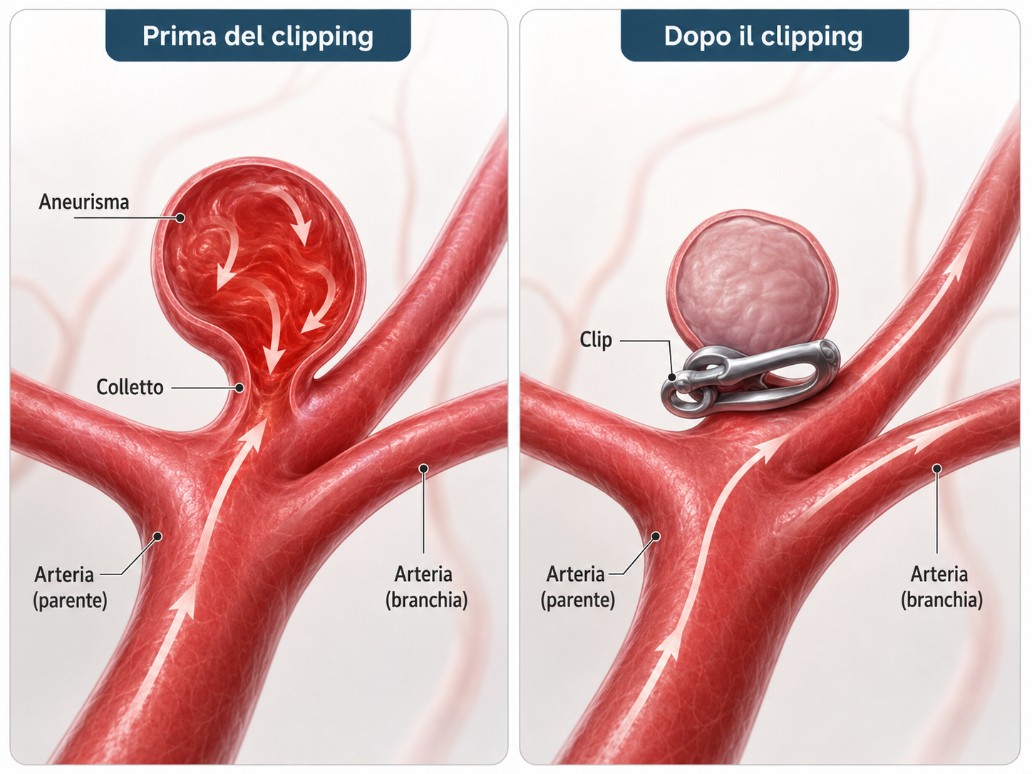
Microsurgical clipping is a vascular neurosurgical procedure used to exclude an aneurysm from the cerebral circulation by placing a small metallic clip across its neck.
The procedure is performed with the assistance of an operating microscope and intraoperative technologies that make it possible to verify complete aneurysm closure while preserving blood flow in the surrounding arteries.
When is clipping indicated?
The choice between microsurgical clipping, endovascular treatment and radiological surveillance is made individually, taking into account:
- the location, size and shape of the aneurysm;
- the width of its neck;
- the presence of nearby arterial branches or branches arising from the aneurysm;
- the patient’s age and general health;
- whether the aneurysm has ruptured;
- the expected durability and stability of the treatment.
Clipping may be particularly suitable for aneurysms with complex morphology, a wide neck, involvement of arterial branches or anatomical characteristics that are less favourable for endovascular treatment.
In selected cases, it can provide immediate and highly durable exclusion of the aneurysm.
How is the procedure performed?
The operation is performed under general anaesthesia.
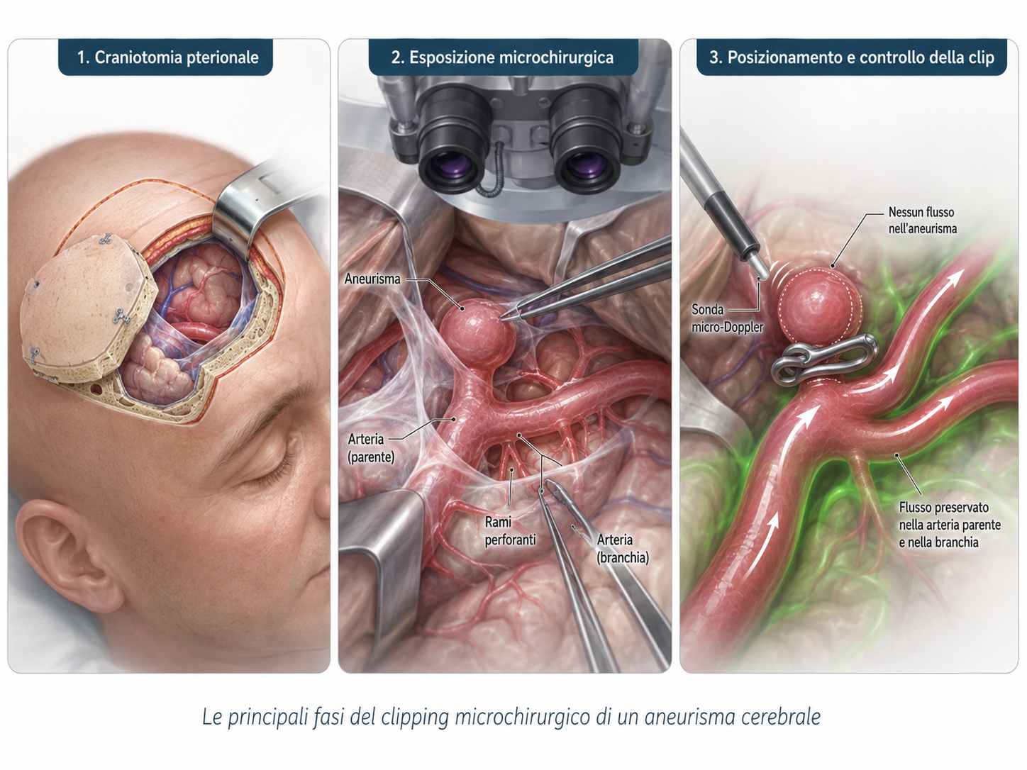
A skin incision is made, followed by a craniotomy, which consists of creating a bone opening to access the aneurysm. For many aneurysms of the anterior cerebral circulation, a frontotemporal approach known as a pterional approach is used.
After opening the dura mater, the procedure continues under microscopic guidance. The neurosurgeon carefully dissects the arachnoid membranes and identifies:
- the artery from which the aneurysm arises;
- the proximal and distal arterial segments;
- the aneurysm neck;
- the surrounding arterial branches and small perforating vessels.
Once the neck has been exposed, a permanent metallic clip is applied. The clip is selected according to the shape and size of the aneurysm.
The objective is to exclude the aneurysm completely while maintaining blood flow through the main artery and its branches.
In more complex aneurysms, several clips may be required, or their position may need to be adjusted until the safest anatomical result is achieved.
Intraoperative checks
The correct position of the clip is verified directly during surgery.
Several techniques may be used.
-
Intraoperative micro-Doppler
A small Doppler probe is used to assess blood flow within the arteries before and after clip placement.
-
Fluorescence angiography
Following the intravenous administration of a fluorescent dye, the operating microscope allows real-time visualisation of blood flow through the cerebral vessels.
This makes it possible to verify:
- complete exclusion of the aneurysm;
- patency of the main artery;
- preservation of the surrounding arterial branches.
The clip can be repositioned immediately when required.
In selected cases, intraoperative neurophysiological monitoring may also be used.
Closure and postoperative care
At the end of the procedure, careful haemostasis is performed. The dura mater is closed, and the bone flap is repositioned and secured with small titanium plates.
Following surgery, the patient is generally monitored for approximately 24 hours in an intensive care unit or specialised monitoring unit.
A brain CT scan with angiographic imaging is then performed to assess:
- correct exclusion of the aneurysm;
- patency of the main cerebral vessels;
- the absence of bleeding or other postoperative complications.
For an unruptured aneurysm and an uncomplicated postoperative course, the hospital stay generally lasts a few days.
During the first few weeks, patients may experience fatigue, moderate headache, tightness around the scar or temporary swelling of the forehead or eyelid. These symptoms generally improve progressively.
What are the risks?
The level of risk depends on the location and complexity of the aneurysm, whether bleeding has already occurred, and the patient’s general health.
Possible complications include:
- bleeding;
- ischaemic stroke;
- temporary or permanent neurological deficits;
- epileptic seizures;
- infection or wound-healing problems;
- hydrocephalus;
- the need for additional procedures;
- anaesthetic or thromboembolic complications;
- in the most severe cases, a risk to life.
Before surgery, these risks are discussed individually and compared with the natural risk of the aneurysm and the available treatment alternatives.
Clipping or endovascular treatment?
Endovascular treatment is performed from within the blood vessels using catheters and may involve coils, stents or flow-diverting devices.
Clipping, by contrast, provides direct visualisation of the aneurysm, the parent artery and its branches. In certain anatomical configurations, it can provide particularly stable long-term closure.
No single technique is best for every patient. Each case should be discussed within a multidisciplinary team to identify the option offering the best balance between safety, effectiveness and durability.
Specialist experience in vascular neurosurgery
PD Dr Thomas Robert completed specialist training in vascular neurosurgery and complex skull base surgery at the Centre hospitalier de l’Université de Montréal – CHUM, under the supervision of Prof. Michel W. Bojanowski.
This experience enabled him to develop specific expertise in microsurgical treatment of cerebral aneurysms, vascular control techniques and skull base approaches.
Dr Robert and Dr Sara Bonasia have extensive experience in the assessment and treatment of cerebral aneurysms, with a combined experience of more than 200 aneurysm clipping procedures.
Procedures are performed at Clinique La Source in Lausanne, in a specialised setting equipped with the technologies required for modern vascular neurosurgery.
A personalised assessment
Every aneurysm has different characteristics.
A specialist consultation allows for detailed analysis of the imaging, comparison of the available treatment options and definition of a personalised strategy.
Would you like a specialist opinion?
We offer a personalised neurosurgical assessment, including a review of your symptoms and brain imaging. You may contact us via the online form or by email : segretariato@neurochirurgia-robert.ch