
Microvascular decompression of the trigeminal nerve
Relieving the nerve while preserving its function
Microvascular decompression of the trigeminal nerve is a neurosurgical procedure indicated for the treatment of trigeminal neuralgia when the pain is caused by contact between the trigeminal nerve and a blood vessel and medical treatment no longer provides satisfactory symptom control or causes significant side effects.
Unlike procedures that deliberately injure part of the trigeminal nerve in order to interrupt pain transmission, microvascular decompression aims to treat the underlying cause of the neurovascular conflict while preserving the integrity and function of the nerve.
For further information about the condition, its symptoms, diagnosis and the different treatment options, patients can consult Our dedicated page on trigeminal neuralgia.
The principle of microvascular decompression
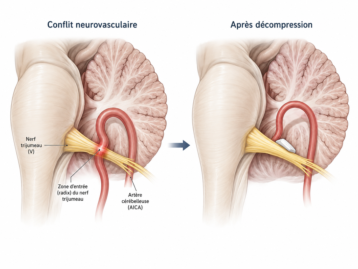
In many patients with classical trigeminal neuralgia, a small artery – most commonly a cerebellar artery – or, less frequently, a vein comes into contact with the trigeminal nerve close to its emergence from the brainstem.
Repeated pulsation of the vessel may affect the nerve at its entry zone and contribute to the characteristic electric shock-like facial pain.
The aim of surgery is to precisely identify the vessel responsible for the conflict, gently separate it from the trigeminal nerve and keep it permanently away from the nerve while avoiding any direct injury to the nerve fibres.
The trigeminal nerve is therefore neither divided nor intentionally damaged.
How the procedure is performed
The procedure is performed under general anaesthesia, with the patient positioned to allow safe access to the posterior part of the skull.
Retrosigmoid approach
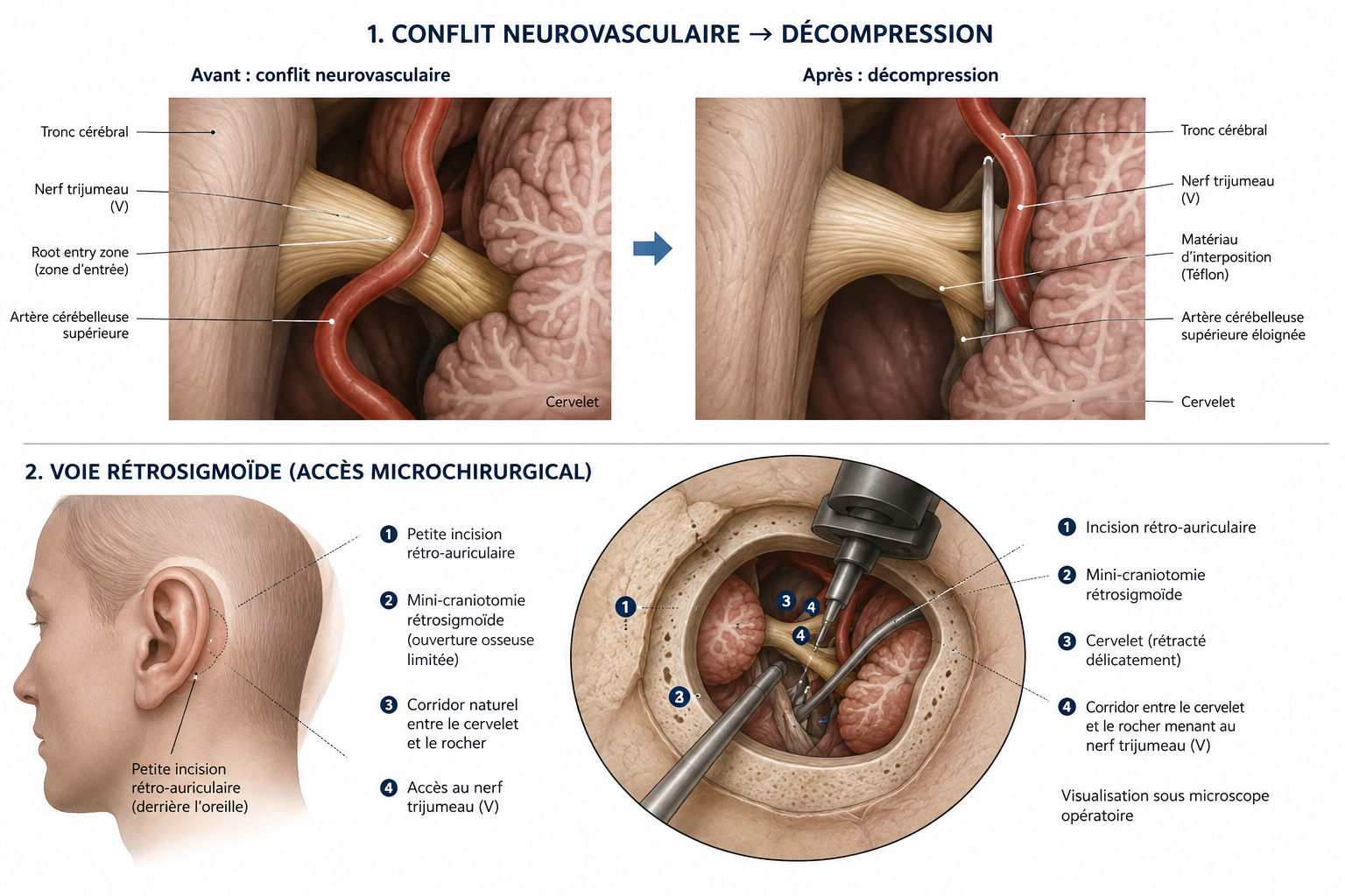
A small skin incision is made behind the ear on the side affected by the neuralgia.
Through a small retrosigmoid bony opening, the neurosurgeon gains access to the posterior cranial fossa.
After opening the dura, a small amount of cerebrospinal fluid is gradually released. This allows the cerebellum to relax naturally and makes it possible to reach the trigeminal nerve while minimising the need for cerebellar retraction.
Microsurgical exploration of the trigeminal nerve
The procedure is performed under an operating microscope using microsurgical techniques.
By following the natural anatomical spaces between the cerebellum and the brainstem, the trigeminal nerve is identified and its course is carefully explored, particularly at the root entry zone, where the nerve emerges from the brainstem.
This is a crucial step because the conflict may be caused by an artery, a vein or, in some cases, several vascular structures at the same time.
Identification of the neurovascular conflict
Once the responsible vessel has been identified, it is gently separated from the nerve.
Particular attention is paid to exploring the entire cisternal course of the trigeminal nerve rather than limiting the assessment to the first visible point of contact.
The objective is to achieve complete decompression of the nerve while preserving the surrounding neurological and vascular structures.
Decompression of the nerve
Different microsurgical strategies can be used depending on the anatomy of the neurovascular conflict.
One option is interposition, in which a small biocompatible material is placed between the vessel and the nerve so that vascular pulsations are no longer transmitted to the trigeminal nerve.
When the anatomy allows it, the vessel may instead be mobilised and transposed, moving it away from the nerve and maintaining it in a position that provides lasting relief from the conflict.
The surgical technique is therefore tailored during the operation to the individual anatomy of the patient and to the type of neurovascular conflict identified.
Precision surgery
Microvascular decompression is performed in a particularly delicate anatomical region containing the brainstem, cerebellum, several cranial nerves and important vascular structures.
For this reason, We use a microsurgical technique focused on the maximum preservation of neurological and vascular structures.
Intraoperative neurophysiological monitoring may also be used, particularly to monitor the auditory pathways and, when appropriate, other relevant cranial nerves, allowing their function to be assessed throughout the different stages of the operation.
Closure of the procedure
Once complete decompression of the trigeminal nerve has been confirmed and no further significant vascular conflicts are identified, the dura is carefully closed.
Particular attention is paid to obtaining a watertight closure in order to reduce the risk of cerebrospinal fluid leakage.
The bony opening is then reconstructed and the surgical wound is closed.
After surgery
In many patients, trigeminal pain disappears immediately after surgery. In others, improvement may occur progressively over the following days.
During the hospital stay, We pay particular attention to any persistence of painful attacks, facial sensation, facial nerve function, hearing, balance and appropriate healing of the surgical wound.
Medication previously used to control trigeminal neuralgia may then be progressively reduced according to the patient’s clinical evolution.
The length of hospital stay depends on the patient’s general condition and postoperative recovery.
What are the advantages of microvascular decompression?
When a neurovascular conflict is responsible for trigeminal neuralgia, microvascular decompression is the surgical treatment that acts directly on the anatomical mechanism responsible for the pain.
Its main advantage is the possibility of achieving long-lasting control of the neuralgia without intentionally damaging the trigeminal nerve.
For this reason, it is particularly considered in patients in good general health whose clinical history and magnetic resonance imaging are compatible with a neurovascular conflict.
Risks of the procedure
As with any intracranial neurosurgical procedure, microvascular decompression carries certain risks.
Possible complications include temporary or permanent hearing impairment, dizziness or balance disorders, changes in facial sensation, facial nerve dysfunction, cerebrospinal fluid leakage, infection, haemorrhage, vascular or neurological complications and, in some cases, persistence or recurrence of trigeminal neuralgia.
Major neurological complications are rare, but the specific risks must always be discussed individually with the patient before surgery.
Our experience
Microvascular decompression of the trigeminal nerve is part of the cranial neurosurgical activity of Our team.
We place particular emphasis on the correct selection of the surgical indication through careful analysis of the clinical history, the characteristics of the pain and the magnetic resonance imaging findings.
During surgery, particular attention is paid to complete identification of the neurovascular conflict, effective decompression of the trigeminal nerve and preservation of the surrounding neurological and vascular structures.
The aim is to provide effective and durable treatment of the neuralgia while preserving trigeminal nerve function as far as possible.
Surgery in Lausanne and follow-up in Ticino
Microvascular decompression procedures for trigeminal neuralgia are performed at the Clinique de La Source in Lausanne, where We have access to the infrastructure and technologies required for microsurgical cranial neurosurgery, including the operating microscope and intraoperative neurophysiological monitoring.
After discharge, patients can be followed directly by Our team in Ticino for postoperative consultations and long-term follow-up.
This allows surgery to be performed in a centre equipped with the technologies required for cranial neurosurgery while providing local postoperative care, ensuring continuity from the preoperative assessment and surgical procedure through to subsequent follow-up.
If you need a specialist evaluation and wish to have it performed with us, you may contact us via the online form or by email : segretariato@neurochirurgia-robert.ch