
Endoscopic third ventriculostomy
Endoscopic third ventriculostomy is a minimally invasive procedure used to treat selected forms of obstructive hydrocephalus. It consists of creating a new pathway for cerebrospinal fluid circulation within the brain, allowing the fluid to bypass the obstruction responsible for its accumulation.
When is this procedure indicated?
This technique is not suitable for every form of hydrocephalus. It is mainly considered when an obstruction prevents cerebrospinal fluid from circulating normally, particularly in cases of aqueductal stenosis, certain tumours, cysts, or malformations of the cerebrospinal fluid pathways.
The indication is established after careful assessment of the patient’s symptoms and brain imaging. In forms of hydrocephalus mainly related to impaired fluid absorption, a ventriculoperitoneal shunt may remain the more appropriate option.
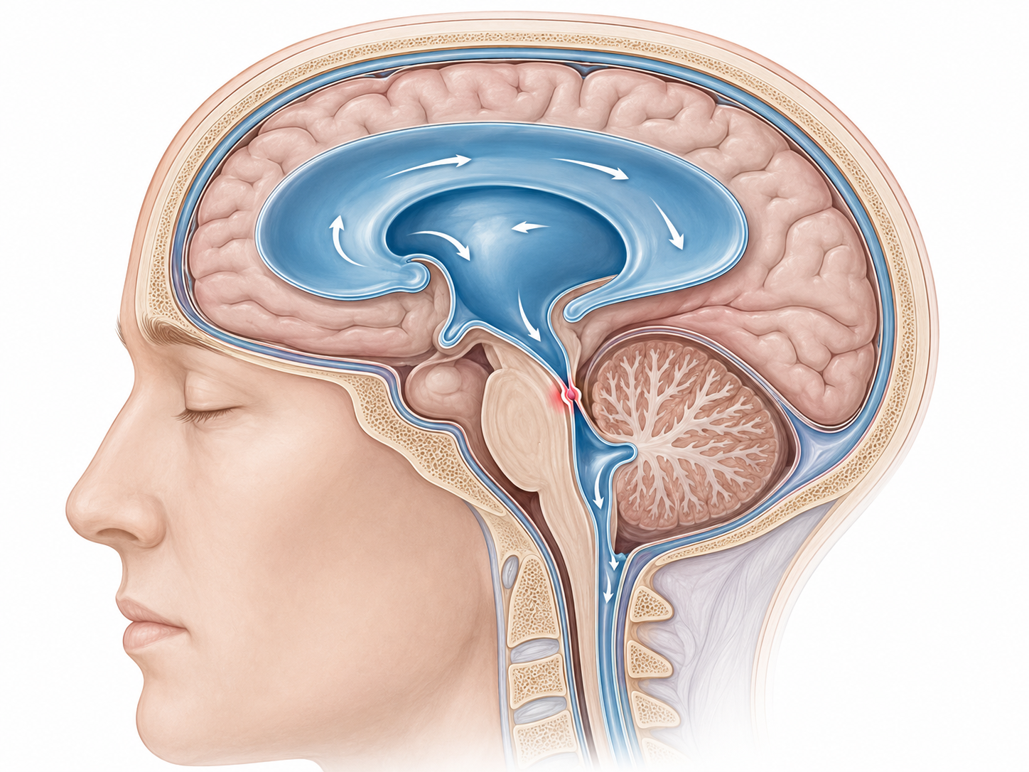
Illustration showing a ctri-ventricular hydrocephalus due to acqueductal stenosis
How is the procedure performed?
The operation is carried out under general anaesthesia.
After a short frontal incision, the neurosurgeon makes a small opening in the skull. An endoscope equipped with a camera is then introduced into the cerebral ventricles.
Under direct visual control, a small opening is created in the floor of the third ventricle. This allows the cerebrospinal fluid to bypass the obstruction and reach the natural fluid spaces at the base of the brain.
The procedure is performed through a limited surgical approach, without the need for a large opening of the skull.
What are the advantages?
When this technique is feasible, its main advantage is that it may avoid the implantation of a permanent shunt system, including a valve and an abdominal catheter.
This reduces the risk of complications related to implanted hardware, such as infection, obstruction, disconnection, or valve malfunction.
Postoperative recovery is generally rapid, and the scar remains small and discreet.
What are the limitations?
The opening created during surgery may occasionally be insufficient or may close again over time. Hydrocephalus may therefore persist or recur, requiring another procedure or the placement of a ventriculoperitoneal shunt.
As with any neurosurgical procedure, endoscopic third ventriculostomy also involves risks, including bleeding, infection, neurological injury, or persistent hydrocephalus. These complications are uncommon but are explained in detail during the preoperative consultation.
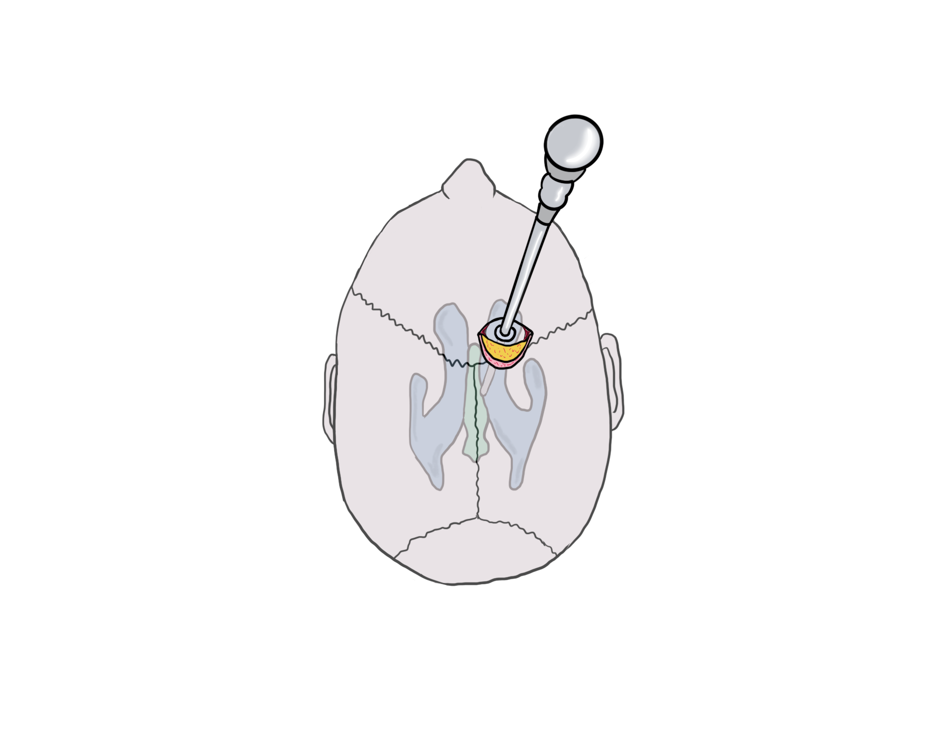
Illustration showing the frontal approach for a ventricular endoscopy
After the procedure
After surgery, the patient is monitored in a neurosurgical unit. Follow-up imaging may be performed depending on the clinical situation.
The hospital stay is generally short, although it depends on the cause of the hydrocephalus and the patient’s overall condition.
The recurrence of severe headaches, vomiting, unusual drowsiness, balance problems, or impaired alertness should lead to prompt medical assessment.
Our approach
Our management is based on a precise analysis of the mechanism of hydrocephalus and a careful selection of patients.
Our experience in cranial neurosurgery and endoscopic surgery allows us to propose an individualised strategy, choosing between endoscopic third ventriculostomy, ventriculoperitoneal shunting, or treatment of the underlying cause of the obstruction.
Our aim is to favour, whenever possible, a technique that is minimally invasive, effective, and adapted to each patient’s anatomy.
Would you like a specialist opinion?
We offer a personalised neurosurgical assessment, including a review of your symptoms and brain imaging. You may contact us via the online form or by email : segretariato@neurochirurgia-robert.ch