Minimally invasive posterior cervical stabilization with CT navigation
Stabilizing the cervical spine with maximum precision
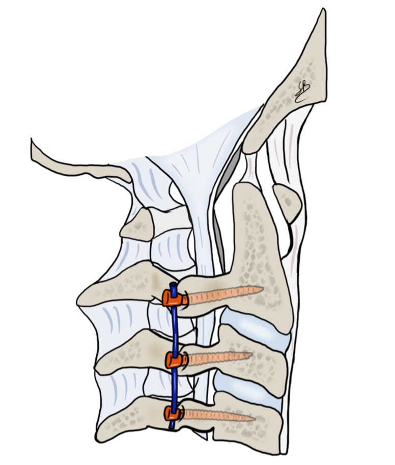
Posterior cervical stabilization is a surgical technique used to restore the stability of the cervical spine by placing screws and rods into the vertebrae.
The procedure is indicated in selected cases of cervical instability, which may be degenerative, traumatic or, less commonly, related to tumor pathology. When neural structures are compressed, stabilization may be combined during the same operation with spinal cord decompression or a posterior cervical foraminotomy.
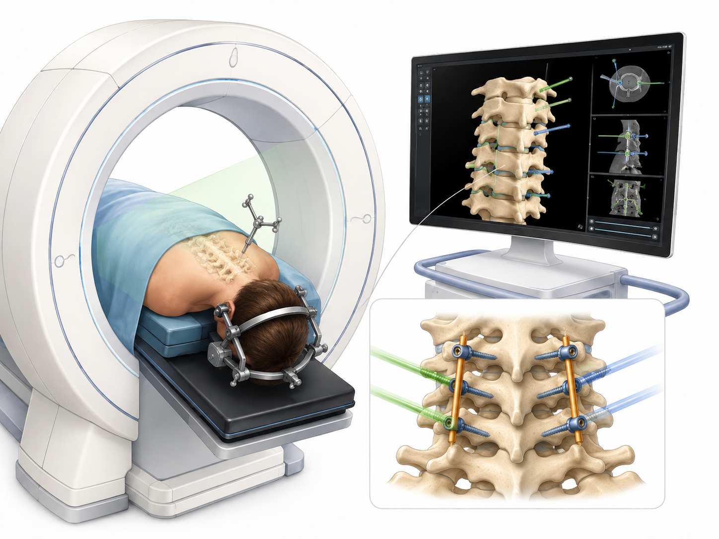
At our center, we perform this procedure using a minimally invasive technique with three-dimensional navigation based on intraoperative O-Arm CT imaging, allowing us to plan and control the placement of the stabilization hardware with a very high degree of precision.
When is it indicated?
Posterior cervical stabilization may be proposed when it is necessary to correct or prevent abnormal movement between two or more cervical vertebrae.
The main indications include:
- Cervical vertebral instability;
- Spondylolisthesis or advanced degenerative changes;
- Instability following trauma;
- The need for stabilization after spinal cord decompression;
- Cervical stenosis associated with instability;
- Disorders affecting the cervicothoracic junction;
- Less commonly, tumor-related or other conditions compromising spinal stability.
Surgery is generally considered when conservative treatment does not provide satisfactory symptom control or when a neurological deficit is present.
Our minimally invasive surgical technique
The procedure is performed under general anesthesia, with the patient positioned prone.
Once the patient has been positioned correctly, a three-dimensional intraoperative CT scan is acquired using the O-Arm system. The images are integrated into the neuronavigation system, allowing the surgeon to visualize the vertebral anatomy and the trajectory of the surgical instruments in real time.
Screw placement under CT navigation
Screw placement in the cervical spine requires particularly high precision because of the close proximity of the spinal cord, nerve roots and vertebral arteries.
For this reason, we systematically use three-dimensional navigation based on intraoperative CT imaging.
The trajectory of each screw can be planned on the three-dimensional images and controlled during insertion, allowing precise selection of:
- The entry point;
- Screw orientation;
- The appropriate screw length and diameter;
- Its relationship to the spinal canal and surrounding vascular structures.
This technology provides a very high degree of accuracy in the placement of spinal instrumentation, which is particularly important in cervical spine surgery.
At the end of screw placement, an additional intraoperative three-dimensional acquisition may also be performed when indicated, allowing immediate verification of screw position before the procedure is completed.
A minimally invasive approach
Depending on the pathology and the levels to be treated, the operation may be performed through two small lateral posterior cervical incisions or through a single central incision.
When neurological decompression is also required, we use a microsurgical technique with a tubular retractor approximately 14–16 mm in diameter.
Through this approach, under microscopic visualization, a small targeted portion of bone and, when necessary, degenerative arthritic tissue compressing a nerve root or the spinal cord can be removed.
Stabilization and decompression can therefore be performed during the same procedure.
Stabilization and decompression during the same operation
When a nerve root is compressed, stabilization can be combined with a posterior cervical foraminotomy, enlarging the foramen through which the nerve exits the spinal column.
When central stenosis is present, spinal cord decompression may also be performed.
The procedure then has two main goals:
- To release the compressed neural structures;
- To restore adequate stability of the cervical spine.
Why choose a posterior approach?
A posterior approach offers specific advantages in selected pathologies.
It allows multiple cervical levels to be stabilized at the same time without necessarily removing the intervertebral discs.
It is also particularly useful for conditions affecting the lower cervical spine and the cervicothoracic junction, an area that can be more difficult to access through an anterior approach.
The choice between anterior and posterior surgery is individualized according to the patient’s anatomy, the type of instability, the presence of neural compression and the number of levels involved.
Advantages of intraoperative CT navigation
The use of intraoperative O-Arm CT imaging combined with neuronavigation is one of the key elements of our surgical technique.
It allows us to:
- Obtain a three-dimensional reconstruction of the spine directly during surgery;
- Plan the trajectory of each screw individually;
- Visualize the position of the instruments in relation to the vertebrae in real time;
- Increase the accuracy of stabilization hardware placement;
- Reduce reliance on external anatomical landmarks alone;
- Verify the result of the stabilization intraoperatively.
This technology is particularly important in the cervical spine, where the anatomical spaces are small and major neurological and vascular structures lie only a few millimeters from the screw trajectories.
Our experience
Spinal surgery and minimally invasive techniques represent an important part of our neurosurgical activity.
Over the years, we have developed particular experience in the use of spinal navigation and intraoperative O-Arm CT imaging, which we use for procedures in which very precise placement of stabilization hardware is required.
The combination of surgical experience, microsurgery, a minimally invasive approach and intraoperative three-dimensional navigation allows us to tailor the procedure to each patient’s anatomy and to perform even complex cervical stabilizations with a high level of control throughout all stages of surgery.
After surgery
Patients can generally stand up and walk on the evening of surgery.
Postoperative pain is controlled with appropriate medication, and discharge usually takes place after approximately two days in hospital, depending on the extent of the operation and the neurological condition.
Clinical and radiological follow-up is then scheduled to assess:
- Wound healing;
- Progression of pain;
- Neurological recovery;
- Stability and position of the instrumentation;
- The vertebral fusion process.
Radiological follow-up is usually continued for approximately 6–9 months after surgery.
Risks of posterior cervical stabilization
As with any cervical spine procedure, posterior stabilization carries certain risks.
Possible complications include infection, hematoma, cerebrospinal fluid leak, neurological deficit, vascular injury, wound-healing or fusion problems and, more rarely, incorrect positioning or loosening of the stabilization hardware.
Preoperative planning, surgical experience and the use of intraoperative CT navigation allow the most delicate stages of the procedure to be controlled with a high degree of precision.
Each indication is nevertheless assessed individually and discussed in detail with the patient before surgery.
Personalized surgery
Not every cervical instability requires the same type of stabilization.
The decision between an anterior, posterior or combined approach, the number of vertebrae to be stabilized and the need for associated decompression are determined on the basis of the symptoms, neurological examination and imaging findings.
The aim is to provide the required stabilization using the least invasive treatment possible, while preserving anatomical structures as much as possible and achieving effective decompression whenever necessary.
If you need a specialist evaluation and wish to have it performed with us, you may contact us via the online form or by email : segretariato@neurochirurgia-robert.ch