
Lumbar disc herniation surgery
Surgical treatment of lumbar disc herniation is one of the most common procedures for spinal disorders. We have been performing this surgery exclusively using a minimally invasive percutaneous technique for more than 7 years and have experience with more than 1,000 surgical cases.
The procedure is performed under general anesthesia and with antibiotic prophylaxis.
To improve surgical precision and minimize tissue opening, the operation is performed under microscopic visualization. The disc responsible for the herniation is identified using fluoroscopy.
The incision is approximately 2 cm long and horizontal (in contrast to the incision used in the conventional technique) and is slightly positioned on the side of the disc herniation.
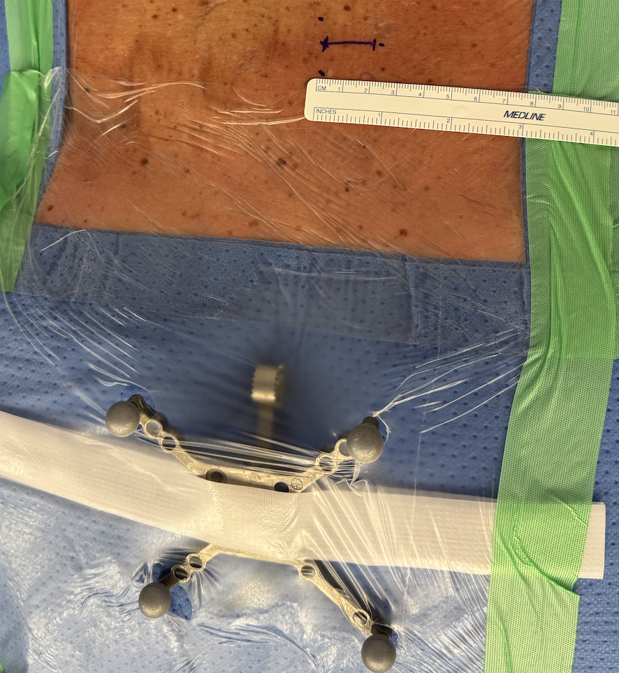
Example of incision size for an herniated disk surgery
After opening the muscular fascia, progressively larger dilators are introduced under navigation guidance until the definitive tubular retractor of 14 mm in diameter is placed.
Inside this tubular retractor, under microscopic visualization, drilling of the vertebral lamina (the bony part of the vertebra) is performed followed by removal of the ligamentum flavum.
After removal of the ligamentum flavum, the dural sac (containing the nerves) and the nerve compressed by the disc herniation become visible.
By gently mobilizing the affected nerve, the disc herniation located under the posterior longitudinal ligament becomes visible. The latter is incised to remove the herniated fragment.
Microdiscectomy, meaning removal of a portion of the disc, is generally not performed.
The retractor is then removed and the muscular fascia and skin are closed after careful hemostasis.
The duration of the procedure is approximately 30 minutes.

Illustration of the tubular retractor for herniated idsk surgery
The minimally invasive technique reduces surgical risks and postoperative pain. However, the risks of surgery cannot be completely eliminated. Our personal statistics for this type of surgery are as follows:
• Postoperative hematoma <0.5%
• Dural tear <0.5%
• Surgical site infection <0.5%
• Direct neurological injury 0%
The overall risk is less than 2% for this procedure.
It is important to understand that the goal of the surgery is to remove the herniated disc and eliminate radicular pain (sciatica). The long-term risk of recurrent disc herniation after this surgery is identical to the risk of developing a new disc herniation without surgery.
The postoperative period includes the following points:
• Hospital stay of 48 hours
• Postoperative physiotherapy after 2 weeks
• No stitches to remove
• Full return to activities after 1 month
If you require an evaluation for an herniated disk and wish to have it performed with us, you may contact us via the online form or by email : segretariato@neurochirurgia-robert.ch