
Ventriculoperitoneal Shunt (VP Shunt)
Surgical treatment of hydrocephalus by cerebrospinal fluid diversion
A ventriculoperitoneal shunt, commonly referred to as a VP shunt, is a neurosurgical technique used to treat certain forms of hydrocephalus, in which an excessive amount of cerebrospinal fluid (CSF) accumulates within the cerebral ventricles.
The aim of the procedure is to restore a proper balance of cerebrospinal fluid circulation, allowing excess fluid to drain from the cerebral ventricles into the abdominal cavity, where it can be normally reabsorbed by the body.
For more information about the condition, the different types of hydrocephalus and their diagnostic assessment, please see our dedicated page on hydrocephalus.
When is a ventriculoperitoneal shunt recommended?
A ventriculoperitoneal shunt is recommended when hydrocephalus is related to impaired circulation or, more commonly, impaired reabsorption of cerebrospinal fluid and cannot be treated by directly eliminating the cause of the obstruction.
One of the most frequent indications in adults is normal pressure hydrocephalus, which mainly affects older patients and may present with:
- Gait and balance disorders;
- Cognitive slowing;
- Urinary disturbances.
Before surgery is recommended, the diagnosis is established on the basis of the symptoms, neurological examination and radiological imaging, particularly brain MRI or CT.
In some patients, additional diagnostic tests, such as a large-volume lumbar puncture, may be necessary to assess the potential response to CSF drainage.
How does a ventriculoperitoneal shunt work?
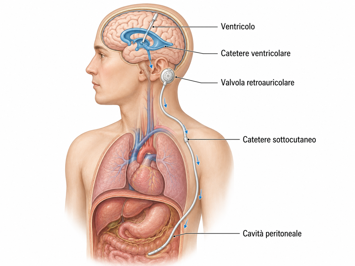
The shunt consists of a system that is positioned entirely under the skin and is therefore not externally visible once the incisions have healed.
The system includes:
- A ventricular catheter, placed within one of the cerebral ventricles;
- A valve, usually positioned under the skin behind the ear;
- A distal catheter, which runs under the skin along the neck and chest to the peritoneal cavity in the abdomen.
Excess CSF is thus drained from the brain into the abdominal cavity, where it is progressively reabsorbed.
The valve makes it possible to control the amount of CSF drained, helping to avoid, as far as possible, insufficient or excessive drainage.
The surgical technique
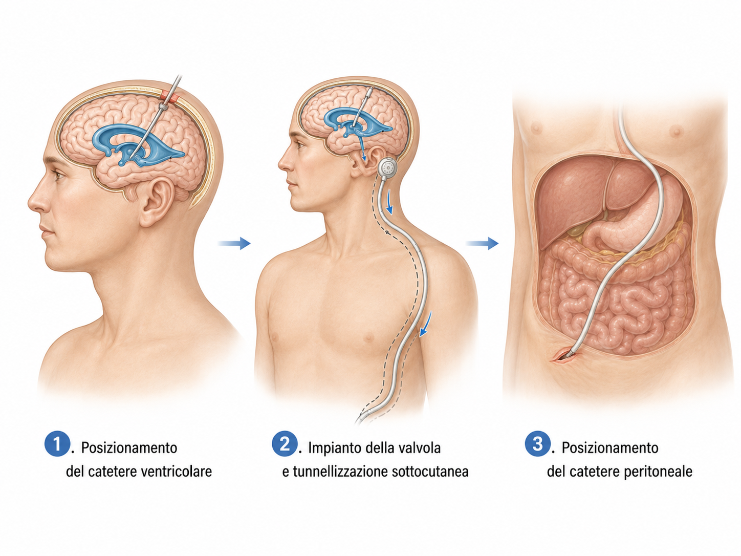
The procedure is performed under general anaesthesia.
Through a small scalp incision, a small opening is made in the skull, through which the ventricular catheter is inserted into the cerebral ventricular system.
The catheter is then connected to the valve positioned under the skin in the retroauricular region.
Using a dedicated instrument, the distal catheter is passed entirely under the skin from the head, along the neck and chest, down to the abdomen.
Through a second small abdominal incision, the distal end of the catheter is finally inserted into the peritoneal cavity, where the cerebrospinal fluid can be naturally reabsorbed by the body.
Before the incisions are closed, the correct functioning of the entire system is verified.
After surgery
After surgery, the patient undergoes neurological and clinical monitoring.
The correct functioning of the shunt, the surgical wounds and the evolution of the symptoms that led to the diagnosis of hydrocephalus are assessed.
Clinical improvement may be gradual. In patients with normal pressure hydrocephalus, one of the first symptoms to improve is often gait quality, while improvement in cognitive or urinary symptoms may take longer.
The shunt is intended to remain implanted long term and therefore requires ongoing neurosurgical follow-up.
Risks and possible complications
As with any neurosurgical procedure, placement of a ventriculoperitoneal shunt carries certain risks.
Complications are generally uncommon, but may include:
- Intracranial bleeding;
- Infection of the shunt or surgical wounds;
- Wound-healing problems;
- Thrombosis or embolism;
- Excessive CSF drainage with possible formation of a subdural haematoma;
- Insufficient drainage or persistence of hydrocephalus;
- Obstruction, disconnection or malfunction of the shunt;
- The need, over time, for partial or complete revision or replacement of the system.
The individual risk is assessed before surgery according to the patient’s age, general medical condition and the cause of the hydrocephalus.
Our experience
We have extensive experience in the diagnosis and surgical treatment of hydrocephalus, including the indication for and placement of ventriculoperitoneal shunts.
Correct patient selection is a fundamental part of treatment, particularly in normal pressure hydrocephalus. For this reason, we place particular importance on the correlation between the clinical presentation, brain imaging and the results of any additional diagnostic tests before recommending surgery.
The objective is not simply to reduce ventricular size, but to achieve a meaningful clinical improvement in the patient’s quality of life.
Surgery in Lausanne and follow-up in Ticino
Ventriculoperitoneal shunt procedures are performed at Clinique de La Source in Lausanne, where we carry out our cranial neurosurgical activity.
After surgery, postoperative consultations and neurosurgical follow-up can also be carried out in Ticino at our practice, allowing patients from Ticino to benefit from continuity of care close to home.
In this way, the entire pathway — from diagnosis and surgical indication through to long-term follow-up — remains under the care of the same neurosurgical team.
If you need a specialist evaluation and wish to have it performed with us, you may contact us via the online form or by email : segretariato@neurochirurgia-robert.ch