Surgical removal of brain tumors
Microsurgery of intracranial lesions
Surgical removal of a brain tumor is one of the main treatment options for intracranial lesions when the tumor’s location, radiological characteristics and the patient’s clinical condition make surgery appropriate.
The goals of surgery are multiple: to obtain an accurate histological diagnosis, reduce the volume of the tumor mass, relieve compression on the brain and, whenever possible, achieve complete removal of the lesion, while preserving brain structures and neurological function as much as possible.
The surgical strategy is tailored individually according to the location and characteristics of the tumor and is discussed in detail with the patient before surgery.
Surgical planning
Before surgery, MRI images and, when required, additional neuroradiological examinations are carefully reviewed.
Planning allows us to select the most direct and least invasive approach possible to the lesion and to identify the brain structures, blood vessels and nerves that must be preserved.
Depending on the location and type of tumor, different supporting technologies may be used:
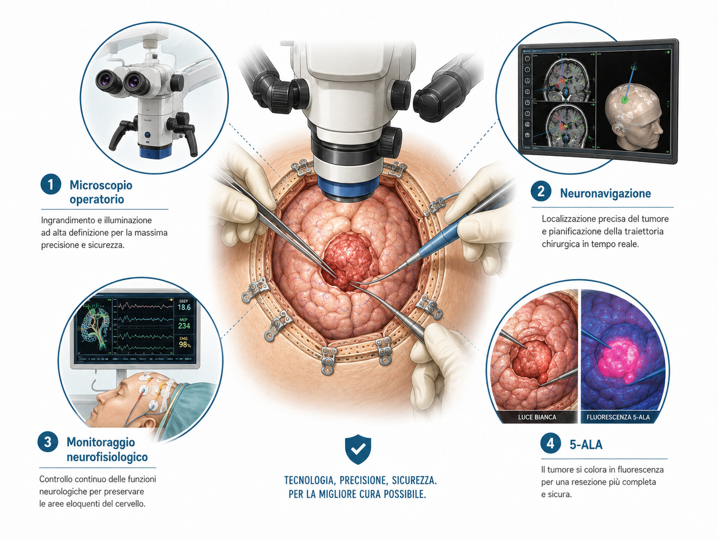
- Neuronavigation, allowing precise localization of the lesion and planning of the surgical trajectory;
- Intraoperative neurophysiological monitoring, particularly when the lesion is located close to functionally important areas of the brain;
- 5-ALA fluorescence, in selected infiltrating tumors, to facilitate intraoperative identification of tumor tissue;
- Intraoperative ultrasound, allowing the location of the lesion and the extent of resection to be assessed during surgery.
How is the operation performed?
The procedure is generally performed under general anesthesia.
The head is stabilized in a dedicated fixation system that allows extremely precise positioning throughout the procedure.
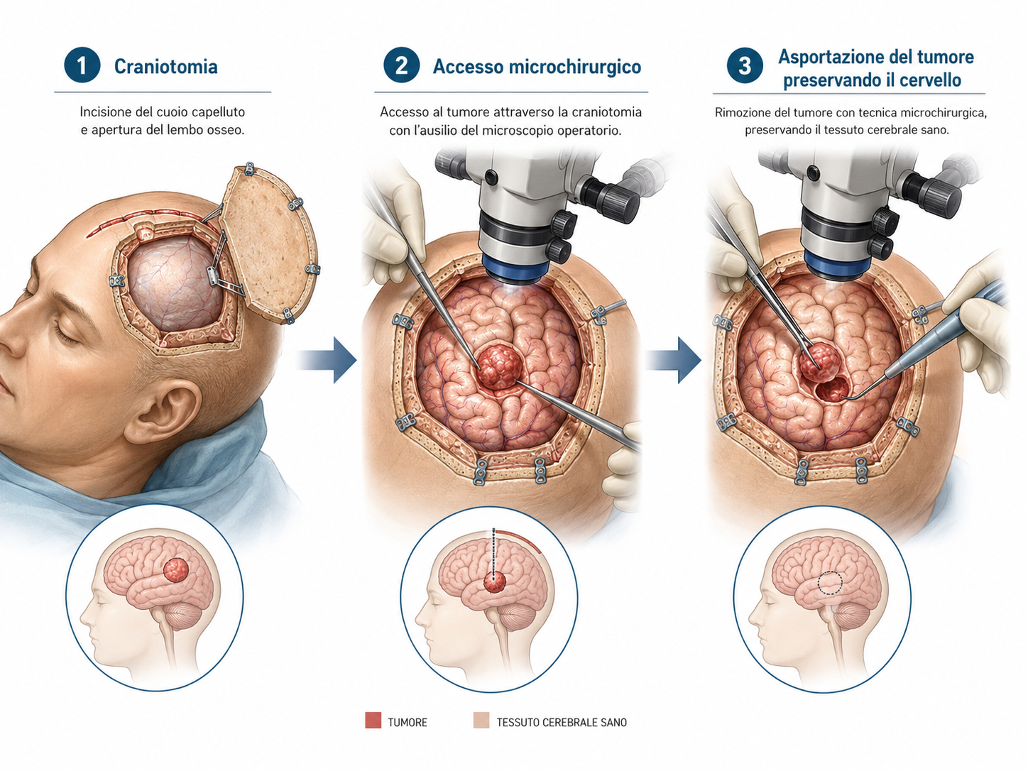
After the skin incision, a craniotomy is performed, meaning a temporary opening of the skull over the area that needs to be reached.
The size and position of the craniotomy are tailored individually, with the aim of obtaining the required surgical access while minimizing tissue trauma.
The procedure is then carried out using a microsurgical technique, with the operating microscope allowing the tumor to be distinguished with high precision from the surrounding neural and vascular structures. Neuronavigation allows precise localization of the lesion. In selected cases, intraoperative neuromonitoring is important to increase safety throughout the procedure.
Removing the tumor while preserving the brain
The fundamental principle of modern brain tumor surgery is to achieve maximal safe resection.
This means removing as much tumor as possible without compromising brain areas that are essential for functions such as movement, sensation, language, comprehension or other higher neurological functions.
For this reason, the final extent of tumor removal can sometimes only be determined during surgery.
In some cases, complete resection can be achieved. In others, when part of the lesion is adherent to or infiltrates particularly important neurological structures, it may be preferable to deliberately leave a small residual component rather than increase the risk of permanent neurological deficit.
The patient’s neurological safety therefore remains the priority over the sole pursuit of radiologically complete resection.
Closing the craniotomy
At the end of the procedure, hemostasis is carefully checked and the dura mater is closed.
The bone flap is then replaced in its original position and secured with small titanium plates and screws, providing immediate stable reconstruction of the skull.
The titanium material can normally remain permanently in place and does not prevent future MRI examinations.
After surgery
At the end of the procedure, the patient undergoes close neurological monitoring.
Depending on the type of surgery and the patient’s clinical condition, the first hours may be spent in an intensive care or continuous monitoring unit.
The following are regularly assessed:
- Level of consciousness;
- Strength and sensation in the limbs;
- Language and comprehension;
- Possible visual disturbances or other neurological deficits;
- Pain, nausea and the condition of the surgical wound.
Mobilization is started progressively as soon as the patient’s clinical condition allows.
Postoperative MRI
After surgery, a postoperative brain MRI is normally performed.
This examination makes it possible to assess the extent of tumor removal, evaluate the surgical result and establish a new baseline scan for subsequent follow-up.
Tumor analysis
The tissue removed during surgery is sent for pathological examination.
This analysis allows precise determination of the nature of the tumor, its biological grade and its molecular characteristics, which are fundamental for establishing prognosis and determining whether further treatment is required.
The final result is generally available after a few days.
Based on the histological findings, the subsequent treatment strategy is then defined. This may involve radiological surveillance alone or, depending on the diagnosis, radiotherapy, chemotherapy or other oncological treatments.
What are the risks?
As with any intracranial neurosurgical procedure, removal of a brain tumor involves certain risks.
The type and magnitude of risk depend mainly on the location, size and nature of the lesion, as well as on the relationship between the tumor and functional brain areas, cranial nerves and blood vessels.
Possible complications include:
- Intracranial bleeding;
- Infection or impaired wound healing;
- Cerebrospinal fluid leakage;
- Thrombosis or embolism;
- Epileptic seizures;
- Hydrocephalus;
- Motor or sensory deficits;
- Language or comprehension disorders;
- Cognitive or behavioral changes;
- The need, in selected cases, for further surgery.
The specific neurological risk is discussed individually before surgery according to the location of the lesion.
Our experience
Surgery for intracranial tumors requires specific training in cranial microsurgical neurosurgery and particular experience in the management of lesions located close to delicate neurological and vascular structures.
PD Dr. Thomas Robert has extensive experience in intracranial surgery and microsurgery, particularly in the treatment of brain tumors and complex skull-base lesions.
The treatment strategy is tailored individually, considering not only the technical possibility of removing the lesion but, above all, the balance between oncological benefit and neurological safety.
Where is surgery performed?
Surgical removal of intracranial tumors is performed at the Clinique de La Source in Lausanne, where the infrastructure required for cranial neurosurgery and perioperative monitoring of these patients is available.
The operation, hospitalization and initial postoperative monitoring are therefore organized in Lausanne.
A care pathway between Lausanne and Ticino
For patients coming from Ticino, care does not end when they are discharged from the clinic.
Once the initial postoperative phase has been completed, subsequent neurosurgical follow-up appointments can also be carried out in Ticino, avoiding repeated travel to Lausanne whenever possible.
Wound assessment, neurological examination, discussion of the pathological results and planning of subsequent imaging follow-up can therefore be integrated into a continuous care pathway between Lausanne and Ticino.
If additional oncological treatments are required, follow-up is coordinated with the specialists involved in the patient’s care.
If you need a specialist evaluation and wish to have it performed with us, you may contact us via the online form or by email : segretariato@neurochirurgia-robert.ch